
- Katalog
- Leistungen
- Über das Unternehmen
- Für Kunden
- Blog
- Kontakt
Sprache und Lieferland
Deutsch · USA
Dermatoscopy is used worldwide as a non-invasive method of examination when skin cancer is suspected, as it significantly increases sensitivity and specificity compared to examination with the naked eye. Dermatoscopy is also known as epiluminescence microscopy (illumination in transmitted light).
To visualize different skin structures for diagnostic purposes, dermatoscopes can be used in contact or non-contact mode, as well as with polarized or non-polarized illumination.
Overall, there are three examination modes with a dermatoscope:
Using different modes and the ability to switch between them can be helpful for the physician when performing differential diagnosis.
In non-polarized contact dermoscopy, the device is pressed directly against the skin. To allow light to penetrate through the stratum corneum and enable assessment of deeper skin layers, an immersion fluid must be applied.
A dermatoscope in its basic configuration consists of focusable magnifying optics, a light source, and a transparent contact plate. Since most of the light hitting the skin is usually reflected, a (glass) contact plate is used, which significantly reduces reflection.
If immersion fluid is applied to the area of skin being examined, the stratum corneum becomes almost transparent, making it possible to assess deeper skin layers. When viewed through magnifying optics, details become visible that are not accessible with the naked eye.
The use of immersion fluid is mandatory in non-polarized contact dermoscopy. The contact plate is fixed on the device.
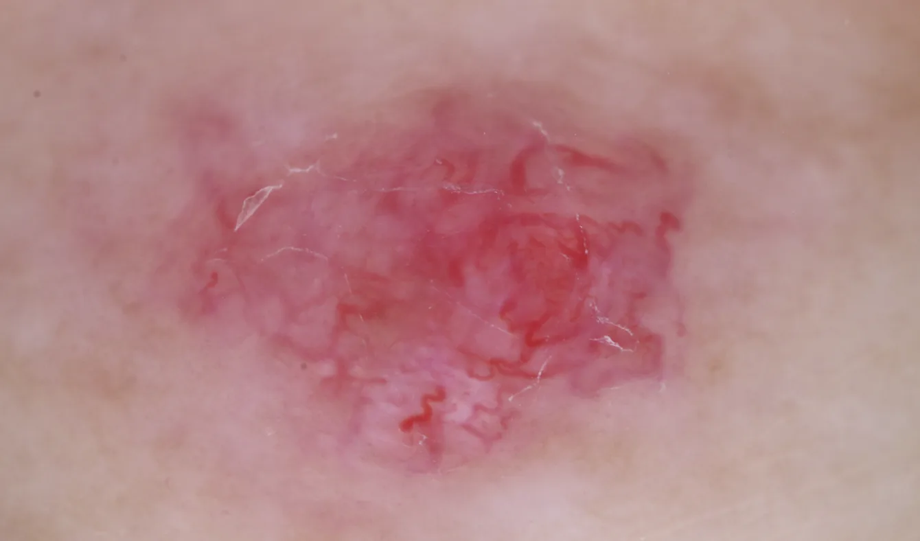 Image with non-polarized illumination
Depending on the type of dermatoscope, the following can be used as immersion fluids:
After the examination, the contact plate must always be wiped with a disinfectant wipe.
Some dermatoscope models come with a small contact plate, which is especially convenient for examining hard-to-reach areas such as interdigital spaces. An alternative in such cases can be polarized non-contact dermoscopy (or the corresponding mode).
more details here
In polarized mode, reflected light from the skin surface is suppressed using a polarization filter. This allows for better visualization of deeper structures of the epidermis.
In this mode, the device is applied to the skin with a contact plate attached. The use of immersion fluid is not mandatory in polarized contact mode.
In both non-polarized and polarized contact dermoscopy, pressing the dermatoscope against the skin exerts pressure on the examined area. On one hand, this may distort the skin’s anatomical geometry, and on the other, it may locally reduce blood flow, making it harder to visualize small vascular structures, for example, in diagnosing basal cell carcinoma.
Therefore, non-contact dermoscopy is increasingly used as an alternative mode.
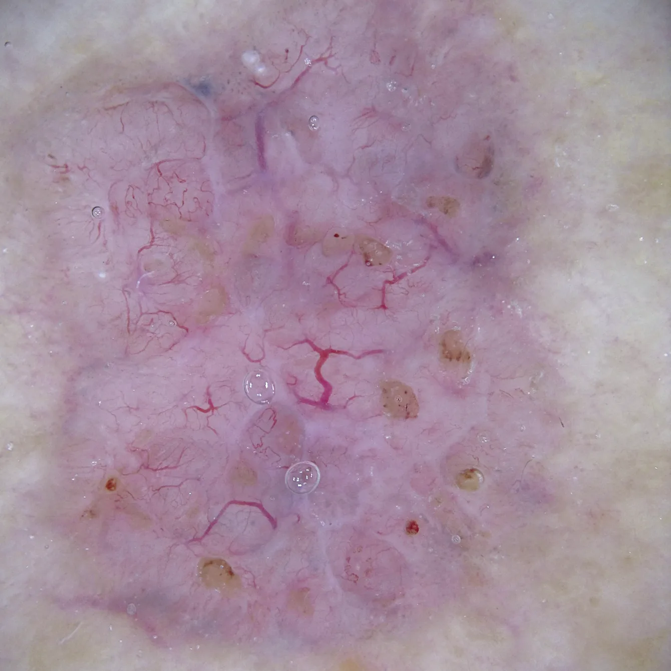 Image with non-polarized illumination
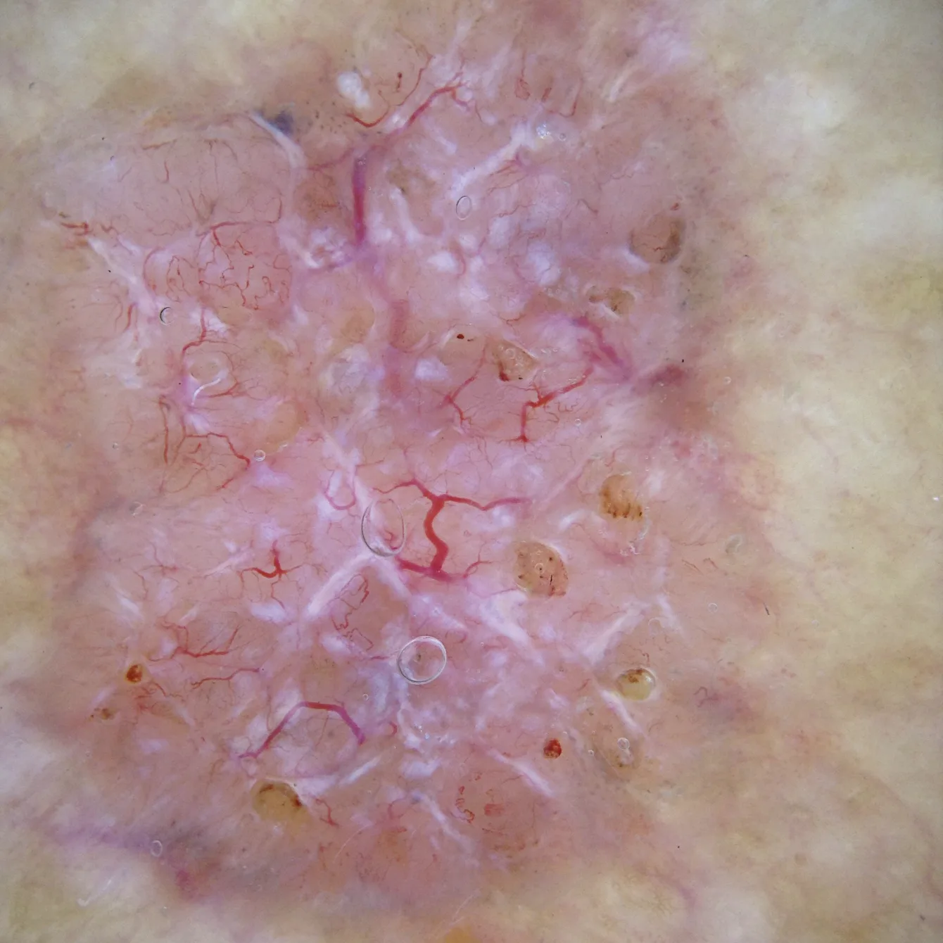 Image with polarized illumination
Using polarized illumination, dermatoscopy can also be performed in non-contact mode. For this, the contact plate of the dermatoscope is removed, and skin areas are examined from a small distance.
This mode is particularly suitable for examining infected skin areas and painful lesions, since no pressure is applied to the dermatoscopic area during non-contact examination.
In this mode, the contact plate is removed from the device. The use of immersion fluid is not only unnecessary but also impossible.
can be viewed here
Alternatively, instead of a contact plate, a diSTANCE working ring can be used, which ensures a constant distance between the skin and the dermatoscope. This allows for examination without pressure, especially when examining raised skin lesions and superficial vessels.
Switching between polarized and non-polarized light helps to more accurately assess skin structures and establish a diagnosis. For example, in models HEINE DELTA 30 PRO, DELTA 30, and DELTAone, a toggle function is available, allowing quick changes in illumination type without replacing the contact plate. Polarized light is activated automatically when the device is switched on.
can be viewed here
Example: melanoma
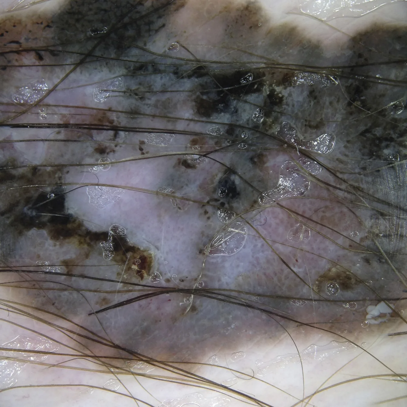 Image with non-polarized illumination
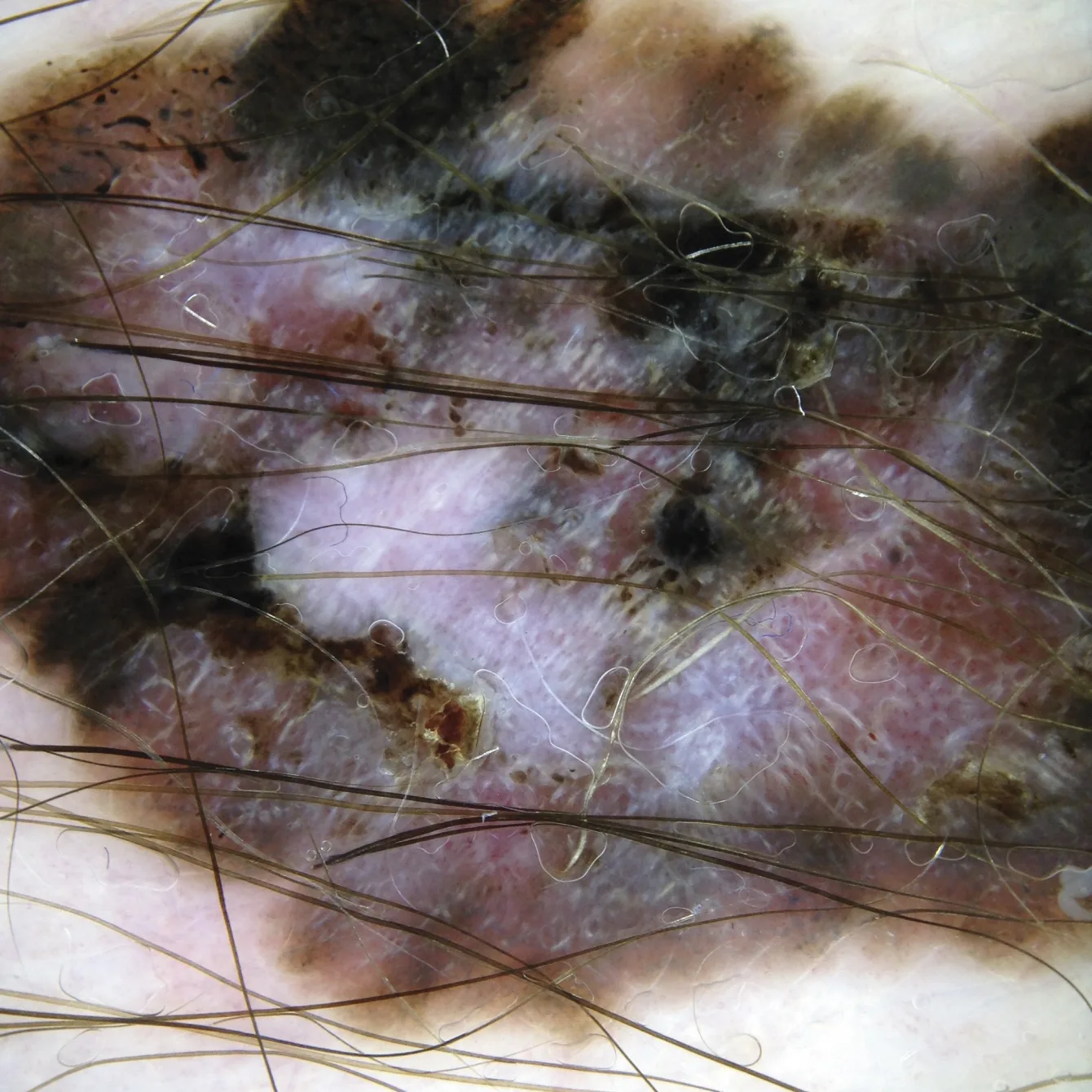 Image with polarized illumination
Modern dermatoscopes allow switching between polarized and non-polarized illumination modes. These two types of illumination visualize different skin structures and thus provide the physician with additional diagnostic information depending on the specific clinical case.
Depending on the depth of their location in the skin, some structures are better visualized with polarization, while others are better without polarization.
Which skin structures are better visualized with non-polarized and polarized contact dermoscopy?
Non-polarized contact dermoscopy
Polarized contact dermoscopy
Superficial structures (from the outer epidermal layer to the dermoepidermal junction).
Deeply located structures, e.g., at the dermoepidermal junction (DEJ) or in the upper dermis.
Superficial whitish structures characteristic of orthokeratosis.
Crystalline and rosette-like structures in skin lesions - so-called shiny white structures associated with collagen and located deeper. Often observed in the diagnosis of basal cell carcinoma.
Blue-white structures in regression areas of malignant melanoma.
In pigmented lesions with melanin at the DEJ or in the dermis (e.g., junctional or blue nevi), darker shades of black and brown are often seen, along with greater variability in the pigment pattern.
Horn pseudocysts or pseudo-follicular openings in seborrheic keratosis.
Vascular patterns and erythema associated with dermal vessels (as examination can be performed without pressure). This can be particularly useful in diagnosing amelanotic or structurally poor melanomas.
TTT HealthCare, HEINE’s primary partner in the countries of the former Soviet Union, organised a two-day educational programme at the HEINE Optotechnik manufacturing facility in Gilching (Munich, Germ
The catalog on the website has already been updated.
The catalog on the website has already been updated.
Produkt wurde in den Warenkorb gelegt
Zum Warenkorb